General
Prevention is better than cure in medicine and it applies in ophthalmology as well. A regular check – up with the ophthalmologist from very early in life can lead to a timely diagnosis and appropriate management for most eye diseases, allowing us to maintain the best possible vision for the rest of our life.
Refractive errors such as astigmatism, myopia and hyperopia, pediatric eye diseases such as squint and amblyopia (lazy eye) and other vision-threatening diseases, such as cataract or glaucoma can be detected at an early stage. Appropriate treatment can improve vision, withhold deterioration with drops, laser treatment or injections depending on the cause of the problem or even fully restore vision with the use of modern and minimally invasive methods.
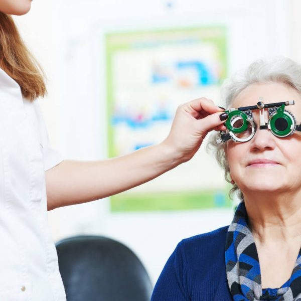
A regular eye check-up by an experienced Ophthalmologist is the first step in order to ensure that your eyesight is and will remain optimum.
According to the international guidelines, adults must check their eyesight every two years.
Respectively, children must undergo an eye check-up when they are born and then: at the first, the third and the fifth year of their life. From the age of 5 and onwards children must undergo an eye check-up every year.
The general check-up for children and adults may include the following:
Refraction – Glasses
Problems of eyesight due to refractive error such as myopia, hyperopia and astigmatism as well as other eye diseases, such as squint, amblyopia (lazy eye) and congenital ptosis may appear in early childhood.
While for adults the treatment of refractive error includes glasses, contact lenses and various laser applications (such as LASIK and PRK) or the placement of special intraocular lenses in front of the crystalline lens of the eye, for children the most appropriate management is prescription of corrective glasses.
The prescription of glasses for children, timely and after a thorough check-up by an experienced Ophthalmologist, has an additional benefit: with the glasses the children not only can see better but their brain learns to see correctly as they grow up and develop physically.
Thus, timely prescription of glasses and eye patching can improve pediatric eye conditions such as lazy eye (or amblyopia) and disorders related to the position of the eyes, such as squint.
Amblyopia and squint are conditions that need to be treated during the first years of a child’s life, before their vision develops fully. The sooner amblyopia and squint are diagnosed and managed the better the outcome for the young person’s quality of life.
Tonometry
Tonometry is a brief and simple eye test that is performed in order to measure the pressure inside the eyes, which is called intraocular pressure.
The eyes contain in the anterior chamber a fluid called the aqueous humor, which is necessary for their nourishment. In healthy eyes, there is constant production of aqueous humor, circulation inside the anterior chamber and drainage in the episcleral vessels. If, for some reason, the drainage of the aqueous humor outside the eye is impeded, then the pressure inside the eye increases.
Increased intraocular pressure is dangerous longterm since it can cause irreversible damage to the optic nerve, which, if not treated timely properly, can lead to progressive loss of visual field and ultimately eyesight due to glaucoma.
Taking intraocular pressure measurements is a significant part of a comprehensive eye examination, as it can be the first indication of glaucoma which is symptom free and therefore can go undetected if not checked.
Tonometry must be performed more often to people having a family history of glaucoma. Since they have a greater risk of developing glaucoma, they need to check their intraocular pressure regularly every year from early adulthood.
This way, glaucoma can be detected timely and managed successfully preventing deterioration of visual field and quality of life.
Phasing
Certain patients that have signs suspicious of glaucoma despite normal intraocular pressure, need to have their pressure checked several times during the day. This process is called phasing.
Gonioscopy
Gonioscopy is an examination that allows the evaluation of the intraocular aqueous drainage system, the so-called anterior chamber angle.
The “angle” refers to the point where the cornea meets the iris and is the point where the aqueous humor is drained outside the eye.
Gonioscopy is used to determine whether the angle is open or closed as well as if there are abnormal blood vessels, adhesions or damage from a previous eye injury.
Angle closure is a condition that may lead to sudden or rapid increase of the intraocular pressure, can be very painful and cause significant visual field loss if it progresses to acute angle closure glaucoma.
Cataract check
Cataract may be evaluated by an Ophthalmologist with a series of tests performed within the framework of a thorough general eye checkup.
The examinations mentioned below help identifying a cataract and evaluating its grade:
- Visual acuity. The visual acuity test measures your vision in different distances. The ophthalmologist will ask you to read letters of different sizes on a board. The examination of visual acuity is an easy, painless and quick way of diagnosing compromised vision due to cloudy intraocular lens that cannot be corrected with glasses
- Slit lamp examination. The slit lamp is a special microscope that allows the examination of the lens of the eye in magnification after instillation of dilating drops in the eye, in order to determine the presence and severity of cataract. Dr. Koumoukeli performs at her practice detailed anterior and posterior segment examination of the eye using the ultramodern KSL – H slit lamp by Keeler. This slit lamp has potential for taking photographs and videos which can help with registration of progressive eye changes and explanation of the signs to the patient.

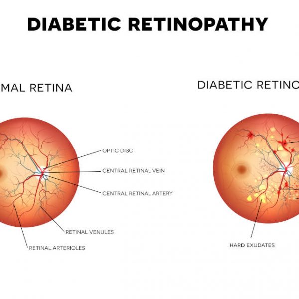
Fundoscopy
Check of the macula, the optic nerve and the periphery of the retina
Fundoscopy is an examination which through a detailed overview of the retinal layer and the vitreous body has the potential to timely diagnose diseases of the part of the eye behind the lens which is called the fundus. Such conditions are diabetic retinopathy, hypertensive retinopathy, macular degeneration, macular hole, epiretinal membrane, glaucoma and others.
Dilation of the pupils is a prerequisite for fundoscopy. After pupil dilation with special drops the Ophthalmologist checks with a special lens the optic nerve, the macula and the vessels at the back of the eye.
The patient will have difficulty seeing properly to drive or read for about four hours after pupil dilation. He is best to avoid driving for several hours after the examination and he needs to wear sunglasses for protecting his eyes from intense sunlight for as long as the pupils remain dilated.
Fundoscopy must be performed every two years to all healthy patients above the age of 60 to check for early signs of macular degeneration.
Patients with diabetes
All patients suffering from diabetes must have their fundus checked once a year. Regular annual fundoscopy is indicated to all diabetics, even those with optimum control of diabetes, as the fluctuations of blood sugar affect the vascular system of the organism, which is depicted clearly in the eye’s fundus.
Stable or no retinopathy on fundoscopy reflects good control of diabetes while new onset vascular changes despite good level of sugar control in the blood checks can help the endocrinologist make appropriate changes to the treatment.