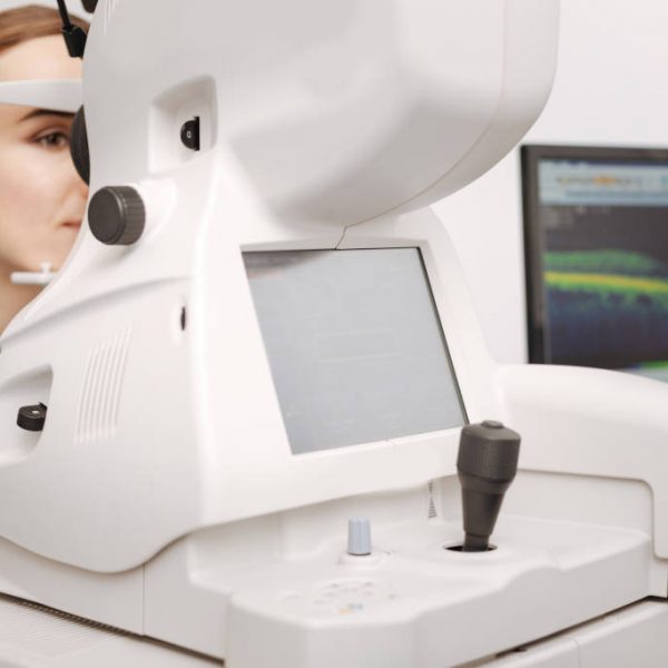
OCT-OPTICAL COHERENCE TOMOGRAPHY
What is optical coherence tomography OCT?
The spectral optical coherence tomography (Spectral Optical Coherence Tomography: OCT-S) is the most modern imaging method of the macula and the optic nerve. It is a cutting-edge technology through which detailed mapping of the posterior eye structures takes place.
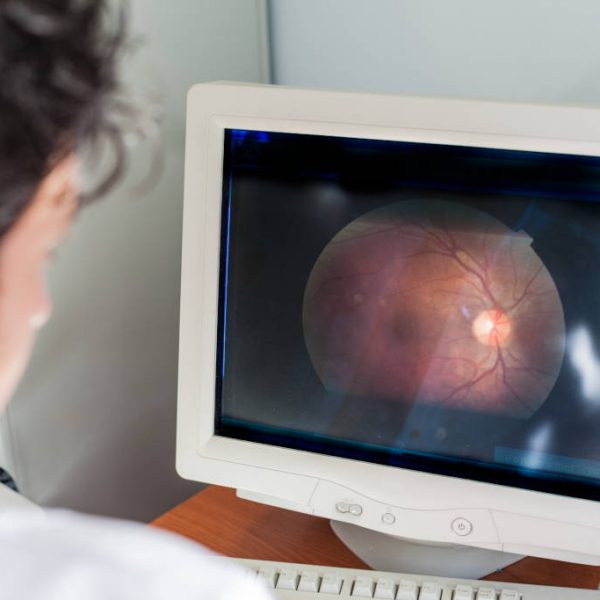
Through the OCT examination, it is accomplished, a full, real time tomography of both the structure and pathology of the suffering area (cases of diabetic retinopathy, age-related macular degeneration, other maculopathy, macular hole, cystoid macular edema, central serous chorioretinopathy, glaucoma, may be easily diagnosed). This diagnostic technique is performed by scanning the point we want to check with a special laser.
The findings are analyzed by the software of the machine and an image that is displayed on the screen is created. This image is a visual intersection of the point of interest, which allows us to identify, diagnose and decide the treatment plan accordingly. In addition to its diagnostic value, OCT is very important for monitoring the evolution of eye diseases (follow-up) and quantifying the therapeutic outcomes.
What is the procedure in the OCT examination (optical coherence tomography)?
The test requires no special preparation from the patient. The patient is placed in front of the machine and within seconds, a detailed recording (mapping) of macula takes place.
OCT ANGIOGRAPHY
The technological revolution is retinal disease diagnosis.
Provides a huge amount of information about various retinal pathology within seconds without the use of contrast media.
As opposed to classic fluorescein angiography, where only the outer capillary network is visible, dye-free OCT – angiography can depict the flow of both the inner and outer retinal capillaries.
Furthermore, the various layers of the retina and the inner choroid as well as their perfusion can be identified separetely, providing important information on a variety of retina-related conditions.
Dye-free OCT angiography has many advantages over classic fluorescein angiography both for the patient and the doctor:
- Being non-invasive examination, it is harmless for the patient as, unlike fluorescein angiography, there is no risk of allergic reaction.
- It can be performed on patients with cardiovascular or renal disorders.
- It does not require the presence of an anesthesiologist.
- It does not require pupil dilation.
OCT – angiography is indispensable for the diagnosis, monitoring and treatment of various retinal conditions, such as:
- Age-related Macular Degeneration
- Retinal vein occlusion
- Diabetic retinopathy and diabetic macular edema
- Macular telangiectasias
- Glaucoma
VISUAL FIELDS
What is a visual field test?
Optical perimetry (visual field test), is the most widespread test of the function of the optic nerve. The technology is based on recognition of a number of light stimuli of various dimensions, which are displayed by the diagnostic system. The examination of the visual fields is paramount for the diagnosis and monitoring of glaucoma, but also in investigating neurological diseases.
The whole process is painless and takes about 15 minutes. It does not require any preparation from the patient (e.g., drops). The examination is actually studying not only the central vision (i.e. how well you see) but also the peripheral vision (i.e. how well we perceive the surrounding environment).
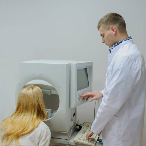
How is the examination of the visual fields performed?
The examination of optical perimetry is performed in a dark room, separately for each eye. The patient’s head is placed in a machine with a large white dome. The patient holds in his hand a special button (joystick) which they press whenever the dome of the machine shows bright light (while they focus on a specific luminous target located just opposite them).
The subject should not move the test eye (not chasing the lights and pressing the button only when they see the light in their peripheral field of vision), otherwise the credibility of the examination is affected. The lights can be strong or weak, in various parts of the dome and some of them check if you are focused. The outcome helps the ophthalmologist in the diagnosis and monitoring of your condition.
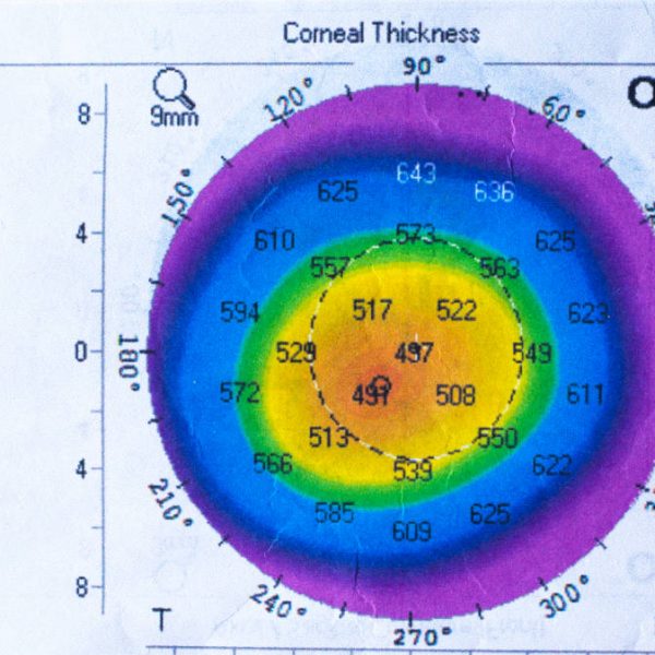
CORNEAL PACHYMETRY
Pachymetry is a special examination measuring the thickness of the cornea.
It plays an important role in evaluating glaucoma as well as in the pre-operative assessment of the patients that undergo refractive surgery with excimer laser.
Pachymetry is performed using an instrument that is called pachymeter. The pachymeter comes in contact with the surface of the cornea and requires the use of local anesthetic drops.
The examination lasts just a few seconds and is totally pain free.
Pachymetry and glaucoma
Several studies have shown that the central thickness of the cornea is a significant indicator for evaluating glaucoma. The combination of increased or even normal intraocular pressure and a thin cornea must be taken into consideration in order to establish the diagnosis and plan treatment for glaucoma.
Pachymetry and refractive surgery
Pachymetry of the cornea is a necessary step of the preoperative assessment for the patient who will undergo refractive surgery with excimer laser.
The outcome of the Pachymetry in combination with the degree of refractive error that needs to be corrected as well as the morphology of the cornea (corneal topography) are the basic factors that will determine the technique that will be used for the operation and the suitability of the candidate.
The thickness of the remaining layer of the cornea is an important factor for refractive surgery.