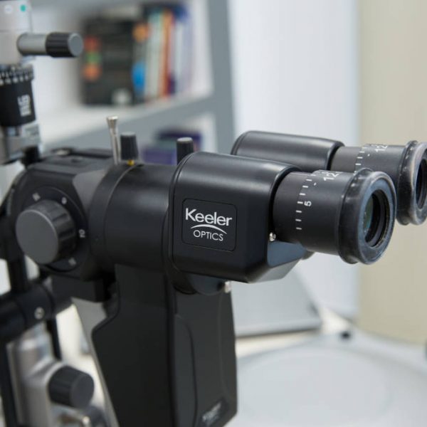
KSL – H Slit Lamp of Keeler
With impressive imaging potential
The ultramodern KSL – H Slit Lamp of the British company Keeler is a diagnostic superpower as it combines exceptional optics with impressive digital imaging potential. With KSL – H, the Ophthalmologist can capture and download photographs or videos of high definition and keep a digital history for each patient. For example, the evolution of the course of treatment of a corneal abrasion or ulcer can be recorded digitally in different stages from the day of first diagnosis to the final day of complete resolution for monitoring purposes.
The Ophthalmologist compares the images or the videos and checks the degree of progress of the disease until resolution. In addition, these digital data can be given to the patient in case hospitalization or referral to a different specialist is considered appropriate.
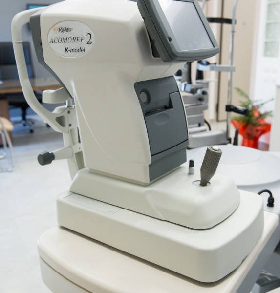
ACOMOREF 2 - Autorefractometer/Keratometer by Righton
Unique in Greece
The right prescription for glasses or contact lenses requires accurate and reliable measurement of the refractive error. This is accomplished with the aid of ACOMOREF 2 by the Japanese company Righton, an ultramodern and unique in Greece autorefractometer/Keratometer with sophisticated functions. It is especially designed for performing difficult measurements in the increasing incidence of eyesight problems secondary to eye fatigue because of the extensive exposure to digital media and the ageing of the population. In addition, it is particularly efficient in the detection and precise measurement of presbyopia at an early stage as well as in the evaluation of refractive problems of various causes.
The biggest advantage of ACOMOREF 2 is the evaluation of accommodation. Sometimes, the patient may have very intense accommodation, meaning that the muscle which moves the lens of the eye is excessively tense. When there is excessive contraction of this muscle that goes undetected by a conventional autorefractometer the prescription of the glasses might be wrong and the effect on eye fatigue significant.
However, ACOMOREF 2 informs the Ophthalmologist that there is intense accommodative spasm in order for him to perform a different type of measurement and therefore obtain an accurate prescription.
Having an even greater speed of measurement and being suitable for even the smallest size of pupil, ACOMOREF 2 guarantees maximum accuracy of measurements, correct spectacle prescriptions and optimum application of toric and multifocal contact lenses.
EAL-100 Focimeter by Righton
Top measurements
The focimeter is an instrument that allows the verification of the prescription of the existing pair of glasses of the patient and it is used by Ophthalmologists before an ophthalmological examination to check the last prescription given to the patient.
With the guarantee of the top Japanese manufacturer Righton, the evolved EAL-100 focimeter achieves maximum measurement accuracy for the lenses of the spectacles or contact lenses.
PachPen Pachymeter of Accutome
Maximum reliability and convenience
The pachymeter allows measurement of the thickness of the cornea of the eye. Pachymetry is considered necessary for patients undergoing refractive surgery to exclude keratoconus. It is also routinely used in glaucoma patients for appropriate adjustment of intraocular pressure measurements according to the thickness of their cornea.
PachPen, the latest technology of the American company Accutome is a portable pachymeter which combines maximum reliability and accuracy with convenient use.
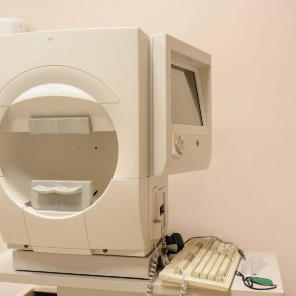
Humphrey HFA – II i Visual Field Tester
The “gold standard” in glaucoma diagnosis and follow up
Humphrey HFA-II i Visual Field Tester is considered to be the “gold standard” in Ophthalmology internationally, providing precision and reliability for visual field assessment. All landmark studies performed internationally on glaucoma have used Humphrey Visual Field Testers for visual field assessment. Thus, the use of Humphrey HFA-II i tester guarantees a friendly and convenient examination for the patient offering at the same time a major advantage to the Ophthalmologist, as the results of the measurements are directly comparable to those of the international bibliography.
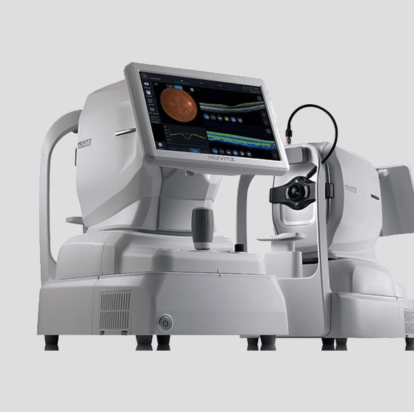
OCT Huvitz HOCT-1/1F
OCT retina, macula, optic nerve head
Fundus Camera
OCT Angiography
The future in technological advancements in fundus imaging is here
This OCT has 3-D auto tracking, auto optimization, auto release, high resolution images, with accurate and stable image averaging. It offers a wide scanning area (12mm x 9mm) and super-fast scanning speed (68000 A-scan/sec) which enhances image resolution and augments accurate diagnosis.
The revolutionary smart Viewing Technology by Huvitz has a Speckle-Noise-Reduction System & Pre-Acquiring Algorithm to acquire high-quality images
There is complete glaucoma analysis (RNFL normative database) which makes it valuable in early diagnosis of glaucoma undetected by visual field testing.
It also provides a 12-megapixel fundus camera with panorama image capture function, extremely useful in diabetic retinopathy screening and optic nerve cupping monitoring in glaucoma.
The Anterior segment module allows measurement and analysis of cornea thickness, angle and 3D image. It helps users work more efficiently by acquiring both anterior and posterior in one place and facilitates the work of every glaucoma specialist.
Finally, this OCT model has the potential for OCT Angiography, a noninvasive technique of visualization of the retina vasculature. It Provides a huge amount of information about various retinal pathology within seconds without the use of contrast media making it ideal for patient comfort without any compromise in resolution or accuracy due to it’s high scanning speed and real time tracking technology.
Dye-free OCT angiography has many advantages over classic fluorescein angiography both for the patient and the doctor:
- Being non-invasive examination, it is harmless for the patient as, unlike fluorescein angiography, there is no risk of allergic reaction.
- It can be performed on patients with cardiovascular or renal disorders.
- It does not require the presence of an anesthesiologist
- It does not require pupil dilation
OCT – angiography is indispensable for the diagnosis, monitoring and treatment of various retinal conditions, such as:
- Age-related Macular Degeneration
- Retinal vein occlusion
- Diabetic retinopathy and diabetic macular edema
- Macular telangiectasias
- Glaucoma
We provide cutting edge technology to support
PREVENTION
IMPROVEMENT
TREATMENT
which are the pillars of our everyday practice
We put the patient in the center of our service