GENERAL
The retinal layer is a thin layer of highly specialized tissue at the back of the eye. The crystalline lens helps the light beams focus onto the retina, which in turn sends the image to the processing areas of the brain through the optic nerve.
The macula is the central portion of the retina, is composed of a thin layer of photosensitive cells and nerve fibers and is responsible for sharp vision (details of objects) and for color vision.
What are the diseases of the retina?
There are several retina conditions that may present with various symptoms and can affect vision significantly either centrally or peripherally. Treating these conditions can, depending on each case, lead to full recovery or mainly slowing down progression of the disease.
Certain diseases of the retina if not treated timely may lead to severe sight impairment.
What are the most common diseases of the retina?
The most common medical retina conditions are the following:
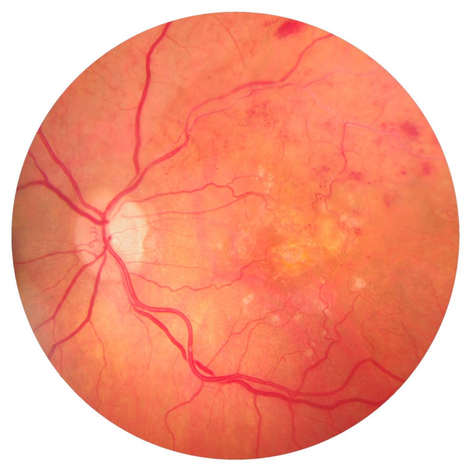
It is noticed to patients with uncontrolled diabetes and it is one of the leading causes of blindness globally. This specific disease is caused by deterioration of the function of the tiny vessels of the retina, which due to diabetes undergo small obstructions and bleedings. As a result, patients may present with macular edema, which causes blurring of central vision.
The macula is a small area at the centre of the retina, which is responsible for detailed vision and color perception. Its degeneration is mainly age related and may be of two types:
A. dry macular degeneration, which presents with slow, gradual changes of the macula cells overtime and is associated with slowly progressive deterioration of central vision and patchy areas in the patient ‘s central visual field
B. wet macular degeneration, which involves neovascularization of the retina and sudden deterioration of the central vision.
Most of them are caused by the development of a thrombosis at a branch or at the central part of a vein (Branch/Central retinal vein occlusion) or artery of the retina (Branch/Central retinal artery occlusion), mostly due to systematic diseases (uncontrolled hypertension, diabetes, cardiovascular diseases). The patients present with bleeding of the retina and deterioration of vision of various levels ,depending on the severity of the episode. These are conditions that need to be addressed immediately and very often require contribution of other specialties, because the cause of the problem lies on general medical conditions rather than the eye itself.
Macular hole is a small defect in the center of the macula at the back part of the eye. It may develop as a consequence of abnormal attachment of the retina on the vitreous body or by some eye injury and presents with central vision defect.
The inner part of our eye is filled with a transparent substance, like a jelly (the vitreous body), which as time goes by becomes less consistent and can get detached from its base away from the retina. Sometimes, this abrupt movement of the vitreous can cause a tear (rupture) of the retina manifesting with photopsias, flashing lights and many black spots that were not evident before the episode.
Retinal detachment occurs when through an untreated retinal tear liquid from the vitreous body passes and builds up underneath the tear. The liquid lifts the retina and subsequently detaches it from the underlying layer of tissue causing significant damage to the retina and threat to the vision if it remains untreated.
Symptoms
What are the symptoms of the diseases of the retina?
Most of the diseases of the retina have some common symptoms, like:
- Photopsias, black spots, flashing lights
- Blurry or distorted vision (metamorphopsia)
- Peripheral vision problems
- Scotomata (small areas of patchy vision centrally or peripherally)
It is important to identify any of the above symptoms and ask for advice from your ophthalmologist.
Causes
What causes the retina diseases?
- Degeneration due to ageing of the retinal cells (macular degeneration etc.)
- Hereditary causes (retinitis pigmentosa)
- Eye injury (macular hole)
- Other diseases such as diabetes (diabetic retinopathy), cardiovascular diseases, hypertension(hypertensive retinopathy), high cholesterol, blood stream diseases
Treatment
How can the diseases of the retina be treated?
Depending on the type of the retinal disease, the aim of the treatment might be improvement or full restoration of vision to its normal levels or mainly prevention of deterioration of vision and preservation of the current levels of eyesight. In some cases, the damage to the patient’s vision is irreversible and the treatment focuses on the slowing down the disease progression and preventing further deterioration of the eyesight.
That is why early diagnosis of retina diseases is crucial.
Treatment of the retina diseases includes various therapeutic choices, depending on the type, the extent and the seriousness of the disease, such as:
1. Laser applications.
The use of therapeutic lasers is often selected for treating macular edema secondary to diabetic maculopathy or retinal vein occlusions. In some cases and depending on the underlying condition, retinal laser treatment can be applied for shrinking and destroying abnormal new retinal vessels that bleed or are at risk of bleeding inside the eye, for example in cases of proliferative diabetic retinopathy or neovascular glaucoma.
Laser retinopexy is also used for delimitating retinal breaks or holes and preventing them from extending and causing retinal detachment.
2. Injectable intraocular administration of anti-VEGF agents.
The injectable administration of medicines in the vitreous body (intravitreal injections) is often selected for the management of conditions such as wet macular degeneration, diabetic retinopathy or retinal vein occlusions in combination with laser applications.
3. Vitrectomy.
It is a microsurgical intervention that involves removing the vitreous body and replacing it by a clear solution. This technique is selected for treating diseases such as retinal detachment, proliferative diabetic retinopathy, macular hole and eye trauma.


